Note : Covid positive reports will be shared as per the local municipal corporation guidelines
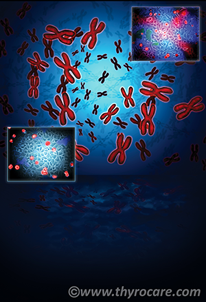
With DNA being considered the blue print of life, every biological function is well coded in our genes. This DNA which dictates every clinical activity within the body is packed in chromosomes within every cell. Every healthy human being is born with a definite number of chromosomes i.e. 22 pairs of autosomes and 1 pair of sex chromosomes. This totals to the presence of 46 chromosomes in genetically healthy individuals
Normal Males - 44 autosomes + X Y Sex chromosomes.
Normal Females - 44 autosomes + X X Sex chromosomes.
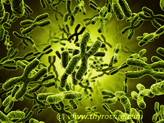
Cytogenetics refers to the study of number and structure of chromosomes within cells. Since chromosomes contain highly coiled DNA, any structural or numerical changes in these directly affect function of genes, resulting in genetic defects/disorders. Cytogenetics is a highly specialized branch of medical science which involves culture of various biological tissues/fluids (peripheral blood, bone marrow, abortus tissue, prenatal tissue), harvesting of cell culture for chromosome preparation, differential stainings (e.g. GTG banding), analysis and reporting of the results. Cytogenetic studies can identify the underlying causes of multiple defects which affect the developing fetus, neonates, children as well as adults. Popularly used techniques under cytogenetics involve Karyotyping and FISH (Fluorescent In-situ Hybridization)
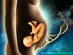
Being the heritable unit of life, DNA on chromosomes become carrier of multiple clinical defects. Though these inherited genetic defects may not be curable, their timely detection can throw light on possibility or highlight the underlying cause for various diseases and disorders. This can aid in taking preventive measures to control the damage and in certain cases appropriate therapeutic intervention can be devised. Since the chromosomes are a constant factor for every human life, cytogenetic testing can be done at any stage of life by both healthy as well as affected individuals. However, few high risk groups are recommended to undergo this test, in case of
Though the above mentioned criteria are to affirmatively undergo a cytogenetic testing, healthy individuals can also be screened to identify presence of any probable underlying disorder.
Since chromosomal analysis is done in living cells, the source can be various biological entities like;
1.Peripheral blood
Cytogenetic testing in peripheral blood sample is generally recommended in adults or neonates to diagnose various syndromes, developmental disorders and chromosome abnormalities (numerical/structural) affecting
fertility.
2.Bone Marrow
Testing of bone marrow is generally recommended for identifying causes of certain cancer types like chronic myelogenous leukemia (CML), acute myelogenous leukemia (ALL), etc. Chromosome analysis in cancer patients
can help in planning treatment, management and studying prognosis of the disease.
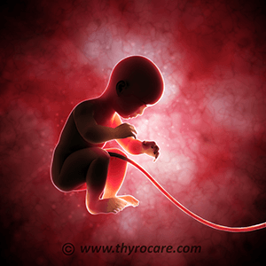
3.Products of conception (POC)
These refer to the tissue formed in the early stages of pregnancy post fertilization including the placenta and aborted fetus. The cytogenetic analysis of the aborted tissue (POC) is recommended if history of more
than two miscarriages, abnormalities on ultrasound prior to pregnancy loss, etc. are observed. This diagnosis can help in recurrence risk estimation and management of future pregnancies.
4.Cord blood
Karyotyping in cord blood involves chromosomal analysis in the fetal blood from the umbilical cord, usually after 20 weeks of pregnancy. This is recommended to diagnose presence of any developmental disorder in
the unborn fetus. In case of third trimester abortion, cord blood can be used for cytogenetic analysis, if available.
5.Chorionic villi
Testing involves analysis in a chorionic villus tissue sample removed from the placenta. This test is generally recommended between the 10th - 12th week of pregnancy and is used to determine
presence of any birth defects in the fetus.
6.Amniotic Fluid
This is present in the amniotic sac of a pregnant woman and is rich in fetal cells. Cytogenetic analysis in amniotic fluid is generally done between the 16th - 20th week of pregnancy and is recommended
in pregnancies presented with USG abnormalities, abnormal maternal serum screening tests and advanced maternal age or bad obstetric history (BOH).
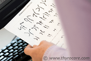
1.Karyotyping
Procedure of arranging the homologous metaphase chromosomes in pairs, from metaphase spreads is called as karyotyping. The method of karyotyping detects chromosome abnormalities bigger than 5 mbp. The whole process
of karyotyping includes setting up of the cell culture (method varies depending on the sample type), arresting the chromosomes in metaphase by addition of spindle inhibitors like colchicine, followed by slide making
and differential staining - banding and finally analysis.
With the availability of automated cytogenetic softwares and advanced microscope systems, the entire karyotyping test can be completed in a shorter time span in comparison to the conventional methodology. The chromosome
spreads on the slides are stained with differential stains and various banding methods are used to compare the band positons with their standard idiograms which increases the accuracy of chromosome identification.
Differential banding techniques used for karyotyping are:
G Banding
The Giemsa banding is the most commonly used and practiced banding technique in diagnostic karyotyping. The method generates dark and light bands and based on the band positions, chromosome identification or karyotyping
is done. The trypsin treated metaphase chromosomes stained with Giemsa, can be easily viewed and analyzed using a light microscope. The dark bands highlight the adenine and thymine rich regions, while the guanine
and cytosine regions are visualized as light bands.
R Banding
R banding is opposite to that of G banding. The guanine-cytosine rich regions produces dark bands whereas the thymidine-adenine rich regions produce light/bright bands. This method uses hot saline to denature AT-rich
DNA and stain the under denatured GC rich regions. R-banding is helpful for analyzing the telomeric ends of the chromosomes, since these areas usually stain light with G-banding.
C Banding
C-banding is used to stain the centromeric chromosome regions. This method also stains the constitutive heterochromatin and secondary constrictions of the chromosomes.
Q Banding
Quinacrine was the first dye to be successfully used for karyotyping chromosomes. This dye binds primarily to the adenine and thymine rich regions of the DNA, highlighted as bright bands, while the dull bands mark
the cytosine and guanine rich regions.
2. Fluorescence In Situ Hybridization (FISH)
FISH is a popularly used molecular cytogenetic technique that involves use of fluorescent labeled probes to analyze the chromosomes in high resolution and provides information in the DNA or gene level. Unlike karyotyping,
which specifically requires metaphase chromosomes, FISH can be done in non-dividing cells as well, and aids in identification of numerical as well as structural abnormalities.
The probes are designed complimentary to the region of interest in the chromosomes and are tagged to fluorescent dyes like biotin, dinitrophenyl, etc. The stained chromosomes are then viewed under fluorescent microscope
for analysis. These FISH probes can also be utilized for multiplex analysis, wherein probes labeled with different fluorophores can be used simultaneously to analyze and detect multiple variations. This technique
is sensitive to detect micro-deletions, duplications, aneuploidies and translocations in chromosome which is a major limitation in other high resolution banding techniques.
IMPORTANT: PCPNDT ACT 1994 should be followed while working with prenatal tissues such as Amniotic Fluid, Chorionic Villi and Cord blood.
Trust Thyrocare - For the wellness and care of yourself and your baby.
Thyrocare is one of the major brand in diagnostics focused on preventive healthcare. With an existing laboratory floor of 2,00,000 sq. ft., accreditations from ISO 9001:2008, CAP (College of American Pathologists) and NABL (National Accreditation Board for Testing and Calibration Laboratories), and state of the art automations and analyzers already supporting the clinical chemistry domain of the laboratory, care has been taken to ensure the same quality of services is replicated in the cytogenetics domain.
Thyrocare with its well established network of franchisees and efficient logistics ensures samples reaching the laboratory floor is processed and reported with a minimum TAT (Turn Around Time). Cytogenetics testing powered by Thyrocare, ensures superior quality chromosomal analysis and detailed reporting by experienced cytogeneticist is provided to every patient as we understand “Timely intervention can aid in crisis alleviation”.